SEVERS - PULL OF TIGHT MUSCLES DAMAGE THE BONE (HEEL)
SALTER HARRIS DISEASE - DAMAGE TO THE EPIPHYSEAL PLATE
AVULSION FRACTURES - TENDONS WORKING TOO HARD THUS INJURING THE ATTACHED BONE.
PARS INTERARTICULARIS FRACTURES - BONE REMODELLING CANNOT COPE WITH THE HEAVY DEMANDS.
SPONDYLOLYSIS - FRACTURE TO THE VERTEBRAE IN BACK
Monday, 12 January 2015

RED BLOOD CELL AND CALCIUM HOMEOSTASIS
LOW BLOOD OXYGEN LEVELS
KIDNEY RELEASED ORYTHROPOEITEN
STIMULATES RED BONE MARROW
CREATES MORE RED BLOOD CELLS
INCREASED OXYGEN CARRYING ABILITY IN RED BLOOD CELLS
CALCIUM
LOW CALCIUM IN BLOOD LEVELS
PARATHYROID GLANDS RELEASE THE PARATHYROID HORMONE
INCREASED LEVELS OF PTH
INCREASES ACTIVITY OF OSTEOCLASTS WHICH RELEASE CALCIUM INTO THE BLOOD.
KIDNEY RELEASED ORYTHROPOEITEN
STIMULATES RED BONE MARROW
CREATES MORE RED BLOOD CELLS
INCREASED OXYGEN CARRYING ABILITY IN RED BLOOD CELLS
CALCIUM
LOW CALCIUM IN BLOOD LEVELS
PARATHYROID GLANDS RELEASE THE PARATHYROID HORMONE
INCREASED LEVELS OF PTH
INCREASES ACTIVITY OF OSTEOCLASTS WHICH RELEASE CALCIUM INTO THE BLOOD.
Q Angles in hip
The Q angles are formed depending on the width of the pelvis and the degree in which the femur sits, A wider pelvis means the femur would articulate at a wider angle, the wider the angle, the more chance the person would suffer from glenu varum. A womens Q angle should be no more than 22 degrees whilst mens is 18 degrees.
Women would have pubic arches greater than 90 degrees.
Women would have pubic arches greater than 90 degrees.
Thursday, 8 January 2015
HIP MUSCLES
GLUTEUS MAXIMUS
GLUTEUS MEDIUS
GLUTEUS MINIMUS
PIRIFORMIS
QUADRATIS FEMORIS
SUPERIOR GEMELLUS
ILLIOPSIS (PSOAS MAJOR AND ILLIACUS)
ADDUCTORS
GLUTEUS MEDIUS
GLUTEUS MINIMUS
PIRIFORMIS
QUADRATIS FEMORIS
SUPERIOR GEMELLUS
ILLIOPSIS (PSOAS MAJOR AND ILLIACUS)
ADDUCTORS
MUSCLES OF THE LEG
QUADRICEP
VASTUS RECTUS (RECTUS FEMORIS)
VASTUS MEDIALIS
VASTUS LATERALIS
VASTUS INTERMEDIALUS
HAMSTRING
SEMITENDINOSUS
BICEPS FEMORIS
SEMIMEMBRANOSUS
LOWER LEG - ANTERIOR
TIBIALIS ANTERIOR
LOWER LEG - POSTERIOR
GASTROSCEMIUS
SOLEUS
VASTUS RECTUS (RECTUS FEMORIS)
VASTUS MEDIALIS
VASTUS LATERALIS
VASTUS INTERMEDIALUS
HAMSTRING
SEMITENDINOSUS
BICEPS FEMORIS
SEMIMEMBRANOSUS
LOWER LEG - ANTERIOR
TIBIALIS ANTERIOR
LOWER LEG - POSTERIOR
GASTROSCEMIUS
SOLEUS
Wednesday, 31 December 2014
BREAKDOWN OF MUSCLE
WHOLE MUSCLE - MUSCLE IS COVERED BY EPIMYSIUM
FASCICULUS - FASCICULUS IS COVERED BY PERIMYSIUM
MUSCLE FIBER - A SINGLE MUSCLE FIBER IS COVERED BY ENDOMYSIUM
MYOFIBRIL:
SARCOLEMMA: Aids the transmission of action potential from the motor neuron to the muscle fibers.
ACTIN: Found within the sacromere on the z disk which create the sliding filaments when connected to the myosin.
SATELLITE CELLS: Facilitate growth, development and adaptation to injury, immobilisation and training of the muscle fibers.
SARCOPLASMIC RETICULUM: Loops around the myofibril and stores calcium which is need to create sliding filaments.
MYOSIN: Two protein strands twisted together one end folded into a globular head
TRANSVERSE TUBULES: Allows waste products to move in and out of the myofibril, makes the myofibril respon to sarcolemma developments.
FASCICULUS - FASCICULUS IS COVERED BY PERIMYSIUM
MUSCLE FIBER - A SINGLE MUSCLE FIBER IS COVERED BY ENDOMYSIUM
MYOFIBRIL:
SARCOLEMMA: Aids the transmission of action potential from the motor neuron to the muscle fibers.
ACTIN: Found within the sacromere on the z disk which create the sliding filaments when connected to the myosin.
SATELLITE CELLS: Facilitate growth, development and adaptation to injury, immobilisation and training of the muscle fibers.
MYOSIN: Two protein strands twisted together one end folded into a globular head
TRANSVERSE TUBULES: Allows waste products to move in and out of the myofibril, makes the myofibril respon to sarcolemma developments.
Monday, 8 December 2014
THE MOVEMENT OF BLOOD THROUGH THE HEART
- Deoxygenated blood arrives via the superior and inferior vena cava.
- The blood moves into the right atrium and passes through the tricuspid valve.
- The blood then moves into the right ventricle which then passes through the pulmonary valve.
- Blood then arrives in the pulmonary artery which carries the deoxygenated blood to the lungs.
- The lungs cause gaseous exchange to occur which removes the carbon dioxide from the blood and refuels it with oxygen.
- The newly oxygen rich blood then returns to the heart via the pulmonary vein.
- The blood passes into the right atrium and through the mitral valve.
- Blood arrives in the right ventricle and then passes through the aortic valve.
- Once through the aortic valve, the oxygen rich blood arrives in the aorta which delivers the blood to the organs and tissues through arteries.
- Once the oxygen rich blood has been sapped by the organs and tissues, the process will start over again and the veins will carry the deoxygenated blood back to the heart.
BLUE = DEOXYGENATED BLOOD
RED = OXYGENATED BLOOD
ITALICS = SYSTEMIC CIRCULATION
NORMAL = PULMONARY CIRCULATION
VEINS, ARTERIES & CAPILLARIES
VEINS = BRINGING DEOXYGENATED BLOOD BACK TO THE HEART AND LUNGS
ARTERIES = BRING OXYGENATED BLOOD TOWARDS THE REST OF THE BODY
CAPILLARIES = TINY VESSELS WHICH CONNECT VEINS WITH ARTERIES, FACILITATE GASEOUS EXCHANGE TO OCCUR AND THE REMOVAL OF WASTE PRODUCTS.
CAPILLARIES
Precapillary sphincters: Maintain and controls blood flow from the arterioles into the capillaries.
Thoroughfare channel: Allows blood to freely move through the arteriole to the venule.
Vascular shunt: The distribution of blood to the working muscles around the body.
Metarterioles: Connect the venules and arterioles together.
VEINS AND ARTERIES
Veins carry oxygenated blood from the lungs back into the heart, this is the pulmonary circulation. However in the systemic circulation they carry deoxygenated blood from the rest of the body back to the heart.
Arteries carry deoxgenated blood from the lungs back into the heart, this is the pulmonary circulation. However in the systemic circulation they carry oxygenated blood from the heart to the rest of the body.
COMPOSITION OF VEINS & ARTERIES
Tunica interna - Thin inner layer of the vein and artery.
Tunica media - Thin middle layer of the vein and artery.
Tunica exertina - Thick outer layer of the vein and artery.
Endothelium cells- Layer of cells within the vein and artery.
Valve - controls blood flow within the vein and artery.
Lumen- the space or hole in the vein and artery which allows the flow of blood.
COMPOSITION OF CAPILLARIES
Endothelium cells - the layer of cells within the capillary.
ARTERIES = BRING OXYGENATED BLOOD TOWARDS THE REST OF THE BODY
CAPILLARIES = TINY VESSELS WHICH CONNECT VEINS WITH ARTERIES, FACILITATE GASEOUS EXCHANGE TO OCCUR AND THE REMOVAL OF WASTE PRODUCTS.
CAPILLARIES
Precapillary sphincters: Maintain and controls blood flow from the arterioles into the capillaries.
Thoroughfare channel: Allows blood to freely move through the arteriole to the venule.
Vascular shunt: The distribution of blood to the working muscles around the body.
Metarterioles: Connect the venules and arterioles together.
VEINS AND ARTERIES
Veins carry oxygenated blood from the lungs back into the heart, this is the pulmonary circulation. However in the systemic circulation they carry deoxygenated blood from the rest of the body back to the heart.
Arteries carry deoxgenated blood from the lungs back into the heart, this is the pulmonary circulation. However in the systemic circulation they carry oxygenated blood from the heart to the rest of the body.
COMPOSITION OF VEINS & ARTERIES
Tunica interna - Thin inner layer of the vein and artery.
Tunica media - Thin middle layer of the vein and artery.
Tunica exertina - Thick outer layer of the vein and artery.
Endothelium cells- Layer of cells within the vein and artery.
Valve - controls blood flow within the vein and artery.
Lumen- the space or hole in the vein and artery which allows the flow of blood.
COMPOSITION OF CAPILLARIES
Endothelium cells - the layer of cells within the capillary.
Wednesday, 3 December 2014
SLIDING FILAMENT THEORY
ROLE OF CALCIUM IN THEORY
- When binding with the troponin, calcium changes its shape which in effect removes the blocking of tropmyosin which allows the myosin heads to attach to the actin.
- Calcium levels vary depending on the action - when muscle goes to rest the myosin head needs to deattach from the actin filament therefore calcium levels drop to allow the myosin head to move back to its original position.
SLIDING FILAMENT THEORY
- The sliding filament theory occurs in the sarcomeres, which run laterally along the myofibril.
- Each sarcomere is made up of two filaments, actin and myosin.
- When we decide to move, calcium attaches onto the troponin of the actin, this then changes shape causing topomyosin to move around allowing the myosin head to attach onto the actin.
- The attachment of the myosin head onto the actin is called a 'CROSS BRIDGE'.
- Once the connection is complete, the ATP allows the myosin head to push the actin, this is called the power stroke and causes the sarcomere to shorten.
- To disconnect the cross bridge, ADP allows the myosin head to return to its original position, this is called the recovery stroke.
- If there is still enough calcium the process will keep going until the calcium is transported back into the sarcopaslmic reticulum, therefore causing the muscle to relax.
- When binding with the troponin, calcium changes its shape which in effect removes the blocking of tropmyosin which allows the myosin heads to attach to the actin.
- Calcium levels vary depending on the action - when muscle goes to rest the myosin head needs to deattach from the actin filament therefore calcium levels drop to allow the myosin head to move back to its original position.
SLIDING FILAMENT THEORY
- The sliding filament theory occurs in the sarcomeres, which run laterally along the myofibril.
- Each sarcomere is made up of two filaments, actin and myosin.
- When we decide to move, calcium attaches onto the troponin of the actin, this then changes shape causing topomyosin to move around allowing the myosin head to attach onto the actin.
- The attachment of the myosin head onto the actin is called a 'CROSS BRIDGE'.
- Once the connection is complete, the ATP allows the myosin head to push the actin, this is called the power stroke and causes the sarcomere to shorten.
- To disconnect the cross bridge, ADP allows the myosin head to return to its original position, this is called the recovery stroke.
- If there is still enough calcium the process will keep going until the calcium is transported back into the sarcopaslmic reticulum, therefore causing the muscle to relax.
Tuesday, 2 December 2014
NEUROMUSCULAR CONTROL
ACTION POTENTIAL released into AXON TERMINAL
Provokes SODIUM CHANNELS to open which release sodium particles into the AXON TERMINAL.
This then causes DEPOLARIZATION to occur which causes CALCIUM CHANNELS to open, thus releasing CALCIUM into the AXON TERMINAL.
CALCIUM then binds with VESICLES which then move to the front of the PRE-SYNAPTIC MEMBRANE.
Once the VESICLES reach the PRE-SYNAPTIC MEMBRANE, the vesicles release the CALCIUM into the SYNAPTIC CLEFT.
The Vesicles carrying the CALCIUM and SODIUM cross the SYNAPTIC CLEFT until they reach the ACH RECEPTORS at the muscle fiber front called the MOTOR END PLATE.
This then opens ION CHANNELS within the ACH RECEPTORS and allows both SODIUM and POTASSIUM to pass through.
The SODIUM pass through whilst POTTASIUM moves way from the muscle fiber, thus causing DEPOLARIZATION in the fiber.
DEPOLARIZATION spreads through the muscle fiber and across the sarcolemma.
The DEPOLARIZATION causes CALCIUM to appear in the muscle fiber, which is therefore used for contractions etc.
Provokes SODIUM CHANNELS to open which release sodium particles into the AXON TERMINAL.
This then causes DEPOLARIZATION to occur which causes CALCIUM CHANNELS to open, thus releasing CALCIUM into the AXON TERMINAL.
CALCIUM then binds with VESICLES which then move to the front of the PRE-SYNAPTIC MEMBRANE.
Once the VESICLES reach the PRE-SYNAPTIC MEMBRANE, the vesicles release the CALCIUM into the SYNAPTIC CLEFT.
The Vesicles carrying the CALCIUM and SODIUM cross the SYNAPTIC CLEFT until they reach the ACH RECEPTORS at the muscle fiber front called the MOTOR END PLATE.
This then opens ION CHANNELS within the ACH RECEPTORS and allows both SODIUM and POTASSIUM to pass through.
The SODIUM pass through whilst POTTASIUM moves way from the muscle fiber, thus causing DEPOLARIZATION in the fiber.
DEPOLARIZATION spreads through the muscle fiber and across the sarcolemma.
The DEPOLARIZATION causes CALCIUM to appear in the muscle fiber, which is therefore used for contractions etc.
Sunday, 23 November 2014
MUSCLE CONTROL
TYPES OF CONTRACTIONS
CONCENTRIC: The shortening of a muscle whilst creating tension
ECCENTRIC : The lengthening of a muscle whilst creating tension
ISOMETRIC: Muscle length stays the same whilst creating tension
MUSCLE CONTROL
AGONIST: Known as the prime mover, the agonist is the muscle which creates movement.
ANTAGONIST: Plays the opposite role to the agonist, the antagonist muscle relaxes or opposes the agonist to create a better contraction. It can play different roles, for one it can relax the agonist, secondly it can create some tension to prevent injury or it can contract fully to add precision and control to the movement.
NEUTRALIZER: Prevents any irrelevant action created by the agonist.
FIXATOR: The fixator muscle contracts to keep other areas stable.
SYNERGIST: Known to assist the agonist muscle when contracting.
CONCENTRIC: The shortening of a muscle whilst creating tension
ECCENTRIC : The lengthening of a muscle whilst creating tension
ISOMETRIC: Muscle length stays the same whilst creating tension
MUSCLE CONTROL
AGONIST: Known as the prime mover, the agonist is the muscle which creates movement.
ANTAGONIST: Plays the opposite role to the agonist, the antagonist muscle relaxes or opposes the agonist to create a better contraction. It can play different roles, for one it can relax the agonist, secondly it can create some tension to prevent injury or it can contract fully to add precision and control to the movement.
NEUTRALIZER: Prevents any irrelevant action created by the agonist.
FIXATOR: The fixator muscle contracts to keep other areas stable.
SYNERGIST: Known to assist the agonist muscle when contracting.
TYPES OF JOINTS
Synathrosis - Unmovable joints (fibrous joint)
Diarthrosis - Freely movable joints (synovial joints)
Amphiarthrosis - Slightly movable joints (cartilaginous joints)
SYNATHROSIS JOINTS
These are types of joints which are unmovable due to the fibrous tissue keeping the joint strong, an example would be skull sutures.
DIARTHROSIS JOINTS
These are also known as synovial joints due to the synovial fluid which allows the joint to be able to freely move. There are various different types of synovial joints:
Gliding joint: Allows gliding or sliding of two joints, for example the carpals in the wrist.
Hinge joint: Works similar to a hinge on a door, allows flexion and extension of the joint.
Ball and socket joint: Allow all movement apart from gliding an example would be the shoulder.
Condyloid joint: Allows flexion and extension in one plane whilst adduction and abduction in another, an example would be the index finger.
Saddle joint: Only found in the thumb (carpometacarpal joint) Similar to ball and socket joint however the joint sits over the metacarpal like a saddle.
Pivot joint: Enables the joint to pivot meaning rotation of the joint, for example the neck.
AMPHIARTHROSIS JOINTS
These are slightly moveable joints due to the hyaline cartilage which attaches the joints together. An example would be the pubis symphysis in the hip girdle or the spinal vertebrae.
Diarthrosis - Freely movable joints (synovial joints)
Amphiarthrosis - Slightly movable joints (cartilaginous joints)
SYNATHROSIS JOINTS
These are types of joints which are unmovable due to the fibrous tissue keeping the joint strong, an example would be skull sutures.
DIARTHROSIS JOINTS
These are also known as synovial joints due to the synovial fluid which allows the joint to be able to freely move. There are various different types of synovial joints:
Gliding joint: Allows gliding or sliding of two joints, for example the carpals in the wrist.
Hinge joint: Works similar to a hinge on a door, allows flexion and extension of the joint.
Ball and socket joint: Allow all movement apart from gliding an example would be the shoulder.
Condyloid joint: Allows flexion and extension in one plane whilst adduction and abduction in another, an example would be the index finger.
Saddle joint: Only found in the thumb (carpometacarpal joint) Similar to ball and socket joint however the joint sits over the metacarpal like a saddle.
Pivot joint: Enables the joint to pivot meaning rotation of the joint, for example the neck.
AMPHIARTHROSIS JOINTS
These are slightly moveable joints due to the hyaline cartilage which attaches the joints together. An example would be the pubis symphysis in the hip girdle or the spinal vertebrae.
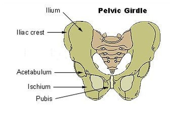
HIP GIRDLE
PARTS OF THE HIP
- ILIAC CREST: The outer crest of the ilium.
- ILIUM: The largest aspect of the hip, the proximal part of the hip.
- ISCHIUM: The bone which hangs down from ilium, houses the obiturator foramen.
- PUBIS: Two bones which are attached together by the pubic symphysis.
- PUBIC SYMPHYSIS: A piece of cartilaginous tissue which connect the two pubis bones together.
- ACETABULUM: The process in which connects the hip to the femur.
- OBITURATOR FORAMEN: The two holes situated in the ischium bone which allow nerves and blood vessels to pass through the hip girdle.

- ILIAC CREST: The outer crest of the ilium.
- ILIUM: The largest aspect of the hip, the proximal part of the hip.
- ISCHIUM: The bone which hangs down from ilium, houses the obiturator foramen.
- PUBIS: Two bones which are attached together by the pubic symphysis.
- PUBIC SYMPHYSIS: A piece of cartilaginous tissue which connect the two pubis bones together.
- ACETABULUM: The process in which connects the hip to the femur.
- OBITURATOR FORAMEN: The two holes situated in the ischium bone which allow nerves and blood vessels to pass through the hip girdle.
VERTEBRAE COLUMN
Parts of the column:
Atlas
Axis
Cervical - 7 vertebrae
Thoracic - 12 vertebrae
Lumbar - 5 vertebrae
Sacrum *
Coccyx *
*= fused together
Ligaments
Anterior longitudinal ligament: Runs up the front of the vertebrae column attaching the vertebraes, it's known to be the stronger ligament out of the two.
Posterior longitudinal ligament: Runs behind the vertebrae column attaching the vertebraes together, it's the weaker and more vulnerable ligament - thinner in lumbar and cervical regions.
 Vertebrae discs
There are 23 discs in the invertebrate column.
Annulus Fibrosis: The wall of the disc, the annulus fibrosis is the thick edge of the disc which keeps the nucleus pulposus from escaping,
Nucleus Pulposus: This makes up 25% of the disc, it's a soft jelly-like substance which sits in between the annulus fibrosis, by age the water content within the pulposus decreases, this is why elderly people begin to gradually 'shrink'. The nucleus' position varies depending on the movement of the spine,
Structure of vertebrae

Vertebrae foramen: This is where the spinal chord is placed.
Body: The body of the vertebrae, where this annulus fibrosis and nucleus pulposus is found. The anterior and posterior longitudinal ligament runs down the body.
Spinous process: We can usually feel this process run down the back of our spine.
Atlas
Axis
Cervical - 7 vertebrae
Thoracic - 12 vertebrae
Lumbar - 5 vertebrae
Sacrum *
Coccyx *
*= fused together
Ligaments
Anterior longitudinal ligament: Runs up the front of the vertebrae column attaching the vertebraes, it's known to be the stronger ligament out of the two.
Posterior longitudinal ligament: Runs behind the vertebrae column attaching the vertebraes together, it's the weaker and more vulnerable ligament - thinner in lumbar and cervical regions.

There are 23 discs in the invertebrate column.
Annulus Fibrosis: The wall of the disc, the annulus fibrosis is the thick edge of the disc which keeps the nucleus pulposus from escaping,
Nucleus Pulposus: This makes up 25% of the disc, it's a soft jelly-like substance which sits in between the annulus fibrosis, by age the water content within the pulposus decreases, this is why elderly people begin to gradually 'shrink'. The nucleus' position varies depending on the movement of the spine,
Structure of vertebrae

Vertebrae foramen: This is where the spinal chord is placed.
Body: The body of the vertebrae, where this annulus fibrosis and nucleus pulposus is found. The anterior and posterior longitudinal ligament runs down the body.
Spinous process: We can usually feel this process run down the back of our spine.
BONES IN FOOT/ANKLE
- NAVICULAR BONE - WHERE ANKLE AND FOOT MEETS.
- LATERAL/MEDIAL MALLEOLOUS - ANKLE BONE WHICH POINTS OUTWARDS
- CUBOID BONE - LATERAL TARSAL BONE
- CUNIFORM BONE - MEDIAL TARSAL BONE
- METATARSALS
- TALUS - ANKLE BONE
- CALCANEUM - HEEL BONE
-TARSALS
-PHELAENGES

- LATERAL/MEDIAL MALLEOLOUS - ANKLE BONE WHICH POINTS OUTWARDS
- CUBOID BONE - LATERAL TARSAL BONE
- CUNIFORM BONE - MEDIAL TARSAL BONE
- METATARSALS
- TALUS - ANKLE BONE
- CALCANEUM - HEEL BONE
-TARSALS
-PHELAENGES

SHOULDER GIRDLE

ACROMIAN PROCESS
This attaches the scapula to the shoulder girdle, the acromian process attaches to the spine of the scapula to help create simultaneous movement in both bones.
GLENOID CAVITY
This is the cavity in which makes up the glenohumeral joint, this is the attachment of the glenoid cavity and the humerus (upper arm).
CLAVICLE
Aka the collarbone, this attaches the shoulder girdle to the manubrium of the sternum. It also attaches to the acromian process thus causing the joint to be named 'Acromioclavicular' joint.
CARACOID PROCESS
A little hook like process which attaches onto the lateral edge of the scapula.
SCAPULA
The scapula is known as a flat bone due to its flat surface shape, there are three parts which make up the scapula, these are called:
- Supraspinous
- Infraspinous
- Spine
SUPRASPINOUS
The supraspinous is situated above the spine of the scapula and makes up the upper part of the scapula.
INFRASPINOUS
The infraspinous is situated below the spine of the scapala and is the lower majority of the bone.
SPINE
As shown on the diagram below, the spine runs inbetween the supraspinous and infraspinous and is attached to the acromian which is situated as part of the shoulder girdle.

- Supraspinous
- Infraspinous
- Spine
SUPRASPINOUS
The supraspinous is situated above the spine of the scapula and makes up the upper part of the scapula.
INFRASPINOUS
The infraspinous is situated below the spine of the scapala and is the lower majority of the bone.
SPINE
As shown on the diagram below, the spine runs inbetween the supraspinous and infraspinous and is attached to the acromian which is situated as part of the shoulder girdle.
BONES: Composition, aspects, types
Bones are the most important structure in the human frame which allows us to create movement, generate and store calcium, withstand significant force and create growth. Below identifies many different aspects of the bone which would need to be accounted for.
TYPES OF TISSUE IN THE BONE:
CORTICAL:
Cortical bone is seen within the diaphysis of the bone as the middle of the bone is the part which withstands the most pressure and would be most vulnerable to fractures. Cortical bone is sometimes known as 'Dense bone' due to the thickness of its structure. The bone has to renew every 10 years to prevent deterioration. The cortical bone is made up of mostly natural compounds such as calcium and collagen.
CANCELLOUS:
Cancellous bone is the complete opposite in terms of structure compared to cortical bone, it is seen to be a spongy bone, thus that term has become a nickname for the bone. The spongy bone has a similar resemble to a honeycomb structure, this allows nerves and blood vessels to passage through the holes in the structure to give the bone the oxygen and nutrients it needs to keep its strength.
ASPECTS OF THE BONE:
EPIPHYSIS:
Seen at the ends of the bone, there are two epiphysis situated on the bone, these are known as the proximal and distal epiphysis. The epiphysis is where cancellous bone would most frequently be found. The epiphysis is made up of the epiphysis plate and line, both of which help promote ossification. The epiphyeal plate is made up of hyaline cartilage and is the part of the bone which grows during childhood, the epiphyeal line shows the 'leftovers' of the epiphyeal plate in adulthood.
DIAPHYSIS:
The diaphysis is situated in the shaft of the bone and contains cortical bone, the most important job this bone is used for is to store yellow bone marrow which in adults stores fat, this can help promote red blood cell production.
TYPES OF BONE:
Long bone: Long bones are the most common types of bone in the body, the length of the bone is significantly bigger than the width of it, thus the name 'long bone'. These bones can be found in fingers and toes (metacarpals/tarsals), radius, ulna, femur, humerus.
Short bone: Short bones consist of a body which is larger in width compared to its length, they promote support and structure of the wrists and the ankles for example, the most common types of short bones would be the carpal and tarsals in the wrists and ankles.
Flat bone: Flat bones are most obviously flat, they're used for protection of vital organs as they're usually rather large and consist of a thick layer of cortical bone. Examples of the flat bone are the scapula, the skull and the hip (illium)
Sesamoid bone: The sesamoid bone is found imbedded between two tendons, these are present most often next to joints as it serves as protection for the tendon which could come into contact with the moving joint. An example is the patella (knee bone)
Irregular bone:The answer is in the title, irregular bones are bones which don't fall into any other category because it's of an irregular shape. Examples include the Sacrum (end of spinal column), Vertabrae found in the spinal column and the mandible (lower jaw)
BONE STRUCTURE #2
Periosteum: It is a double layer membrane which makes up the outer sheet of the bone tissue, it is significant as it is supplied with blood vessels, nerves and lympatic vessels.
Endosteum: This is the inner layer of the bone surface, this is where osteocytes are present meaning that this is where osteoblasts and osteoclasts work in order to maintain appropriate levels of collagen and calcium levels.
BONE CELLS
Both osteoblast and clasts are used prodominantly for ossification (bone remodeling) and can be found in the endosteum (cortical bone).
Osteoclasts: The first stage of bone remodelling occurs when osteoclasts begin to remove and reabsorb bone tissue found in the endosteum, this tissue would have been deprived of collagen meaning it would need to be removed in order for new collagen to be injected into the tissue.
Osteoblasts: The second stage is when the osteblasts move into lay down new bone tissue full of collagen, the new bone tissue would be layed upon the osteocytes which had previously been stripped by the osteoclasts.
INJURY CONCERNS OF THE BONE
Avulsion fractures: Avulsion fractures are commonly caused by stress placed upon a certain bone, this means that the bone remodeling cannot cope with the ongoing demands of the bone wanting to become stronger.
Spondylolysis: This is most often seen in the vertabrae and it's the degeneative osteoartheritis of the spinal vertebrae coming together. It is sometimes referred to as the 'spotty dog' due to its resemblance of a dogs collar.
Osgood Schlatters Disease: A problem which occurs in the knee, it is the pain and swelling of the ligament due to the inaccurate growth of the eiphyseal plate.
Severs disease: A fracture which occurs in the heel bone, it is most commonly seen in children. It is the inflammation and fracture of the calcaneus which is caused by the achillis tendon pulling too hard on the attachment which is situated on the calcaneus.
Salter Harris fracture: This results in a fracture taking place in the epiphyseal plate which will cause deformation of the plate and also causes the the plate to close off preventing any further growth in the epiphysis.
TYPES OF TISSUE IN THE BONE:
CORTICAL:
Cortical bone is seen within the diaphysis of the bone as the middle of the bone is the part which withstands the most pressure and would be most vulnerable to fractures. Cortical bone is sometimes known as 'Dense bone' due to the thickness of its structure. The bone has to renew every 10 years to prevent deterioration. The cortical bone is made up of mostly natural compounds such as calcium and collagen.
CANCELLOUS:
Cancellous bone is the complete opposite in terms of structure compared to cortical bone, it is seen to be a spongy bone, thus that term has become a nickname for the bone. The spongy bone has a similar resemble to a honeycomb structure, this allows nerves and blood vessels to passage through the holes in the structure to give the bone the oxygen and nutrients it needs to keep its strength.
ASPECTS OF THE BONE:
EPIPHYSIS:
Seen at the ends of the bone, there are two epiphysis situated on the bone, these are known as the proximal and distal epiphysis. The epiphysis is where cancellous bone would most frequently be found. The epiphysis is made up of the epiphysis plate and line, both of which help promote ossification. The epiphyeal plate is made up of hyaline cartilage and is the part of the bone which grows during childhood, the epiphyeal line shows the 'leftovers' of the epiphyeal plate in adulthood.
DIAPHYSIS:
The diaphysis is situated in the shaft of the bone and contains cortical bone, the most important job this bone is used for is to store yellow bone marrow which in adults stores fat, this can help promote red blood cell production.
TYPES OF BONE:
Long bone: Long bones are the most common types of bone in the body, the length of the bone is significantly bigger than the width of it, thus the name 'long bone'. These bones can be found in fingers and toes (metacarpals/tarsals), radius, ulna, femur, humerus.
Short bone: Short bones consist of a body which is larger in width compared to its length, they promote support and structure of the wrists and the ankles for example, the most common types of short bones would be the carpal and tarsals in the wrists and ankles.
Flat bone: Flat bones are most obviously flat, they're used for protection of vital organs as they're usually rather large and consist of a thick layer of cortical bone. Examples of the flat bone are the scapula, the skull and the hip (illium)
Sesamoid bone: The sesamoid bone is found imbedded between two tendons, these are present most often next to joints as it serves as protection for the tendon which could come into contact with the moving joint. An example is the patella (knee bone)
Irregular bone:The answer is in the title, irregular bones are bones which don't fall into any other category because it's of an irregular shape. Examples include the Sacrum (end of spinal column), Vertabrae found in the spinal column and the mandible (lower jaw)
BONE STRUCTURE #2
Periosteum: It is a double layer membrane which makes up the outer sheet of the bone tissue, it is significant as it is supplied with blood vessels, nerves and lympatic vessels.
Endosteum: This is the inner layer of the bone surface, this is where osteocytes are present meaning that this is where osteoblasts and osteoclasts work in order to maintain appropriate levels of collagen and calcium levels.
BONE CELLS
Both osteoblast and clasts are used prodominantly for ossification (bone remodeling) and can be found in the endosteum (cortical bone).
Osteoclasts: The first stage of bone remodelling occurs when osteoclasts begin to remove and reabsorb bone tissue found in the endosteum, this tissue would have been deprived of collagen meaning it would need to be removed in order for new collagen to be injected into the tissue.
Osteoblasts: The second stage is when the osteblasts move into lay down new bone tissue full of collagen, the new bone tissue would be layed upon the osteocytes which had previously been stripped by the osteoclasts.
INJURY CONCERNS OF THE BONE
Avulsion fractures: Avulsion fractures are commonly caused by stress placed upon a certain bone, this means that the bone remodeling cannot cope with the ongoing demands of the bone wanting to become stronger.
Spondylolysis: This is most often seen in the vertabrae and it's the degeneative osteoartheritis of the spinal vertebrae coming together. It is sometimes referred to as the 'spotty dog' due to its resemblance of a dogs collar.
Osgood Schlatters Disease: A problem which occurs in the knee, it is the pain and swelling of the ligament due to the inaccurate growth of the eiphyseal plate.
Severs disease: A fracture which occurs in the heel bone, it is most commonly seen in children. It is the inflammation and fracture of the calcaneus which is caused by the achillis tendon pulling too hard on the attachment which is situated on the calcaneus.
Salter Harris fracture: This results in a fracture taking place in the epiphyseal plate which will cause deformation of the plate and also causes the the plate to close off preventing any further growth in the epiphysis.
Subscribe to:
Posts (Atom)