TYPES OF CONTRACTIONS
CONCENTRIC: The shortening of a muscle whilst creating tension
ECCENTRIC : The lengthening of a muscle whilst creating tension
ISOMETRIC: Muscle length stays the same whilst creating tension
MUSCLE CONTROL
AGONIST: Known as the prime mover, the agonist is the muscle which creates movement.
ANTAGONIST: Plays the opposite role to the agonist, the antagonist muscle relaxes or opposes the agonist to create a better contraction. It can play different roles, for one it can relax the agonist, secondly it can create some tension to prevent injury or it can contract fully to add precision and control to the movement.
NEUTRALIZER: Prevents any irrelevant action created by the agonist.
FIXATOR: The fixator muscle contracts to keep other areas stable.
SYNERGIST: Known to assist the agonist muscle when contracting.
Sunday, 23 November 2014

TYPES OF JOINTS
Synathrosis - Unmovable joints (fibrous joint)
Diarthrosis - Freely movable joints (synovial joints)
Amphiarthrosis - Slightly movable joints (cartilaginous joints)
SYNATHROSIS JOINTS
These are types of joints which are unmovable due to the fibrous tissue keeping the joint strong, an example would be skull sutures.
DIARTHROSIS JOINTS
These are also known as synovial joints due to the synovial fluid which allows the joint to be able to freely move. There are various different types of synovial joints:
Gliding joint: Allows gliding or sliding of two joints, for example the carpals in the wrist.
Hinge joint: Works similar to a hinge on a door, allows flexion and extension of the joint.
Ball and socket joint: Allow all movement apart from gliding an example would be the shoulder.
Condyloid joint: Allows flexion and extension in one plane whilst adduction and abduction in another, an example would be the index finger.
Saddle joint: Only found in the thumb (carpometacarpal joint) Similar to ball and socket joint however the joint sits over the metacarpal like a saddle.
Pivot joint: Enables the joint to pivot meaning rotation of the joint, for example the neck.
AMPHIARTHROSIS JOINTS
These are slightly moveable joints due to the hyaline cartilage which attaches the joints together. An example would be the pubis symphysis in the hip girdle or the spinal vertebrae.
Diarthrosis - Freely movable joints (synovial joints)
Amphiarthrosis - Slightly movable joints (cartilaginous joints)
SYNATHROSIS JOINTS
These are types of joints which are unmovable due to the fibrous tissue keeping the joint strong, an example would be skull sutures.
DIARTHROSIS JOINTS
These are also known as synovial joints due to the synovial fluid which allows the joint to be able to freely move. There are various different types of synovial joints:
Gliding joint: Allows gliding or sliding of two joints, for example the carpals in the wrist.
Hinge joint: Works similar to a hinge on a door, allows flexion and extension of the joint.
Ball and socket joint: Allow all movement apart from gliding an example would be the shoulder.
Condyloid joint: Allows flexion and extension in one plane whilst adduction and abduction in another, an example would be the index finger.
Saddle joint: Only found in the thumb (carpometacarpal joint) Similar to ball and socket joint however the joint sits over the metacarpal like a saddle.
Pivot joint: Enables the joint to pivot meaning rotation of the joint, for example the neck.
AMPHIARTHROSIS JOINTS
These are slightly moveable joints due to the hyaline cartilage which attaches the joints together. An example would be the pubis symphysis in the hip girdle or the spinal vertebrae.
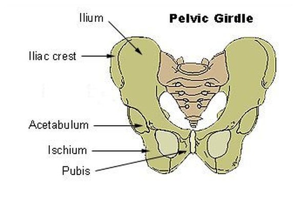
HIP GIRDLE
PARTS OF THE HIP
- ILIAC CREST: The outer crest of the ilium.
- ILIUM: The largest aspect of the hip, the proximal part of the hip.
- ISCHIUM: The bone which hangs down from ilium, houses the obiturator foramen.
- PUBIS: Two bones which are attached together by the pubic symphysis.
- PUBIC SYMPHYSIS: A piece of cartilaginous tissue which connect the two pubis bones together.
- ACETABULUM: The process in which connects the hip to the femur.
- OBITURATOR FORAMEN: The two holes situated in the ischium bone which allow nerves and blood vessels to pass through the hip girdle.

- ILIAC CREST: The outer crest of the ilium.
- ILIUM: The largest aspect of the hip, the proximal part of the hip.
- ISCHIUM: The bone which hangs down from ilium, houses the obiturator foramen.
- PUBIS: Two bones which are attached together by the pubic symphysis.
- PUBIC SYMPHYSIS: A piece of cartilaginous tissue which connect the two pubis bones together.
- ACETABULUM: The process in which connects the hip to the femur.
- OBITURATOR FORAMEN: The two holes situated in the ischium bone which allow nerves and blood vessels to pass through the hip girdle.
VERTEBRAE COLUMN
Parts of the column:
Atlas
Axis
Cervical - 7 vertebrae
Thoracic - 12 vertebrae
Lumbar - 5 vertebrae
Sacrum *
Coccyx *
*= fused together
Ligaments
Anterior longitudinal ligament: Runs up the front of the vertebrae column attaching the vertebraes, it's known to be the stronger ligament out of the two.
Posterior longitudinal ligament: Runs behind the vertebrae column attaching the vertebraes together, it's the weaker and more vulnerable ligament - thinner in lumbar and cervical regions.
 Vertebrae discs
There are 23 discs in the invertebrate column.
Annulus Fibrosis: The wall of the disc, the annulus fibrosis is the thick edge of the disc which keeps the nucleus pulposus from escaping,
Nucleus Pulposus: This makes up 25% of the disc, it's a soft jelly-like substance which sits in between the annulus fibrosis, by age the water content within the pulposus decreases, this is why elderly people begin to gradually 'shrink'. The nucleus' position varies depending on the movement of the spine,
Structure of vertebrae

Vertebrae foramen: This is where the spinal chord is placed.
Body: The body of the vertebrae, where this annulus fibrosis and nucleus pulposus is found. The anterior and posterior longitudinal ligament runs down the body.
Spinous process: We can usually feel this process run down the back of our spine.
Atlas
Axis
Cervical - 7 vertebrae
Thoracic - 12 vertebrae
Lumbar - 5 vertebrae
Sacrum *
Coccyx *
*= fused together
Ligaments
Anterior longitudinal ligament: Runs up the front of the vertebrae column attaching the vertebraes, it's known to be the stronger ligament out of the two.
Posterior longitudinal ligament: Runs behind the vertebrae column attaching the vertebraes together, it's the weaker and more vulnerable ligament - thinner in lumbar and cervical regions.

There are 23 discs in the invertebrate column.
Annulus Fibrosis: The wall of the disc, the annulus fibrosis is the thick edge of the disc which keeps the nucleus pulposus from escaping,
Nucleus Pulposus: This makes up 25% of the disc, it's a soft jelly-like substance which sits in between the annulus fibrosis, by age the water content within the pulposus decreases, this is why elderly people begin to gradually 'shrink'. The nucleus' position varies depending on the movement of the spine,
Structure of vertebrae

Vertebrae foramen: This is where the spinal chord is placed.
Body: The body of the vertebrae, where this annulus fibrosis and nucleus pulposus is found. The anterior and posterior longitudinal ligament runs down the body.
Spinous process: We can usually feel this process run down the back of our spine.
BONES IN FOOT/ANKLE
- NAVICULAR BONE - WHERE ANKLE AND FOOT MEETS.
- LATERAL/MEDIAL MALLEOLOUS - ANKLE BONE WHICH POINTS OUTWARDS
- CUBOID BONE - LATERAL TARSAL BONE
- CUNIFORM BONE - MEDIAL TARSAL BONE
- METATARSALS
- TALUS - ANKLE BONE
- CALCANEUM - HEEL BONE
-TARSALS
-PHELAENGES

- LATERAL/MEDIAL MALLEOLOUS - ANKLE BONE WHICH POINTS OUTWARDS
- CUBOID BONE - LATERAL TARSAL BONE
- CUNIFORM BONE - MEDIAL TARSAL BONE
- METATARSALS
- TALUS - ANKLE BONE
- CALCANEUM - HEEL BONE
-TARSALS
-PHELAENGES

SHOULDER GIRDLE

ACROMIAN PROCESS
This attaches the scapula to the shoulder girdle, the acromian process attaches to the spine of the scapula to help create simultaneous movement in both bones.
GLENOID CAVITY
This is the cavity in which makes up the glenohumeral joint, this is the attachment of the glenoid cavity and the humerus (upper arm).
CLAVICLE
Aka the collarbone, this attaches the shoulder girdle to the manubrium of the sternum. It also attaches to the acromian process thus causing the joint to be named 'Acromioclavicular' joint.
CARACOID PROCESS
A little hook like process which attaches onto the lateral edge of the scapula.
SCAPULA
The scapula is known as a flat bone due to its flat surface shape, there are three parts which make up the scapula, these are called:
- Supraspinous
- Infraspinous
- Spine
SUPRASPINOUS
The supraspinous is situated above the spine of the scapula and makes up the upper part of the scapula.
INFRASPINOUS
The infraspinous is situated below the spine of the scapala and is the lower majority of the bone.
SPINE
As shown on the diagram below, the spine runs inbetween the supraspinous and infraspinous and is attached to the acromian which is situated as part of the shoulder girdle.

- Supraspinous
- Infraspinous
- Spine
SUPRASPINOUS
The supraspinous is situated above the spine of the scapula and makes up the upper part of the scapula.
INFRASPINOUS
The infraspinous is situated below the spine of the scapala and is the lower majority of the bone.
SPINE
As shown on the diagram below, the spine runs inbetween the supraspinous and infraspinous and is attached to the acromian which is situated as part of the shoulder girdle.
Subscribe to:
Posts (Atom)